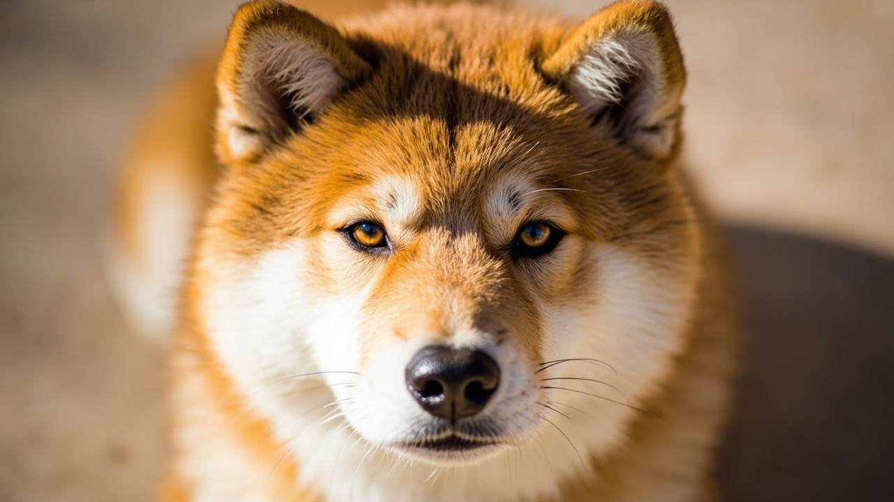
Cherry Eye in Shiba Inus: Causes, Treatment, and Recovery
Cherry eye in a Shiba Inu is a prolapsed third eyelid gland that appears as a red, swollen mass in the inner corner of the eye. It is not painful in itself but requires surgical correction by a veterinary ophthalmologist to prevent long-term dry eye and irritation. Most Shibas recover fully within 2–3 weeks after surgery.
Cherry eye in a Shiba Inu is the common name for prolapse of the nictitating membrane (third eyelid) gland. Instead of sitting quietly behind the lower eyelid, the small tear-producing gland slips out of position and protrudes, forming a smooth, red, cherry-like lump in the inner corner of one or both eyes. Because Shiba Inus have a relatively shallow eye socket and genetically weaker connective tissue anchoring that gland, they are among the breeds predisposed to the condition.
The protrusion blocks normal tear drainage and exposes the gland to air, dust, and friction. If left alone, it can lead to chronic conjunctivitis, corneal ulcers, and reduced tear production (dry eye, or KCS) over the following months and years. That is why veterinary consensus is clear: cherry eye is a surgical condition, not a cosmetic one.
Why Shiba Inus Develop Cherry Eye
The third eyelid gland is held in place by a tiny ligament. In predisposed breeds, this ligament is loose or stretches easily, so the gland pops forward. Contributing factors include:
- Genetics: A congenital weakness in the connective tissue attachment.
- Age: Most cases appear before 2 years of age, often between 6 and 12 months.
- Secondary swelling: Mild conjunctivitis, allergies, or a small scratch can inflame the tissue enough to push an already-loose gland out.
- Breed predisposition: Along with Bulldogs, Cocker Spaniels, Beagles, and Lhasa Apsos, Shibas are over-represented in veterinary ophthalmology caseloads.
Cherry eye is generally not caused by trauma, although a bout of the "Shiba 500" zoomies into a bush can occasionally trigger the first episode in a dog already predisposed.
Recognizing the Symptoms
The visual sign is unmistakable: a pink to deep red, fleshy, oval mass protruding from the corner of the eye nearest the nose. It can affect one eye or both (bilateral cases occur in roughly 30–50% of affected dogs, and the second eye often prolapses within weeks to months).
Other signs to watch for:
- Redness and mild swelling of the conjunctiva
- Excessive tearing or stringy mucus discharge
- Squinting or partial closure of the eye
- Pawing or rubbing at the face
- A dry, dull appearance to the cornea in chronic cases
Because cherry eye can look like a tumor to an untrained eye, prompt veterinary examination is essential to rule out neoplasia (rare in young Shibas) and to plan treatment.
How Vets Treat Cherry Eye in Shibas
1. Surgical Replacement (Preferred)
The gold-standard procedure is the pocket technique (also called imbrication or Morgan pocket). The surgeon creates a small pocket in the connective tissue of the third eyelid, tucks the gland inside, and closes it with fine, absorbable sutures. This preserves 100% of the tear-producing tissue.
A common alternative is the anchoring (tack-down) technique, where the gland is sutured to the periosteum of the orbital rim or the sclera.
Success rates for the pocket technique in published veterinary studies range from 85–95%, with recurrence rates under 10% when performed by a board-certified veterinary ophthalmologist.
2. Gland Removal (Avoided Today)
Older textbooks recommended simply cutting the gland out. This is now considered outdated because removing the gland leaves the dog with a 40–60% lifelong risk of dry eye (keratoconjunctivitis sicca), requiring daily cyclosporine or tacrolimus drops. Shibas already have a higher baseline of immune-mediated eye issues, so preservation is strongly preferred.
3. Conservative Management
In very mild, intermittent cases, a veterinarian may prescribe anti-inflammatory eye drops and an Elizabethan collar to prevent self-trauma while the inflammation settles. This is rarely a permanent fix in Shibas and usually just delays surgery.
What to Expect After Surgery
- Day 1–3: Mild swelling, mild discharge, and some squinting are normal. An E-collar is essential to prevent rubbing.
- Day 7: Recheck visit; sutures are usually dissolving and the gland should be back in position.
- Day 14–21: Full resolution of swelling; most Shibas are back to normal activity.
- Long-term: Monitor the opposite eye, especially in the first year, for bilateral prolapse. Annual eye exams are wise; CHIC testing already requires an OFA eye examination.
Cost and Choosing a Surgeon
In the U.S., cherry eye surgery with the pocket technique typically runs $500–$1,500 per eye at a general practice and $800–$2,000 per eye at a board-certified veterinary ophthalmologist. Although the specialist costs more, the lower recurrence rate and reduced lifetime dry-eye risk make it a worthwhile investment for a breed with a 13–16 year lifespan.
Prevention and Breeding Notes
Cherry eye has a heritable component, so affected Shibas should not be bred. Reputable breeders will discuss any history of the condition and screen breeding stock through organizations like OFA and CHIC. If you are buying a Shiba puppy, ask the breeder directly whether there is any history of cherry eye, luxating patella, or glaucoma in the line.
Acting quickly is the single best thing an owner can do. The longer the gland stays prolapsed, the more it swells and scars, making surgery more difficult and the prognosis less ideal.
FAQ
Can cherry eye in a Shiba Inu go away on its own?
No. Once the gland has prolapsed, it does not return to its normal position without surgical correction. Delaying treatment increases the risk of dry eye and corneal damage.
Is cherry eye painful for a Shiba Inu?
The prolapse itself is not acutely painful, but the resulting irritation, conjunctivitis, and risk of corneal ulceration can become painful and lead to chronic discomfort if untreated.
How long does recovery take after cherry eye surgery in a Shiba?
Most Shibas recover within 2–3 weeks. Swelling and discharge improve noticeably in the first week, and the gland is typically fully repositioned and stable by the third week.
Will my Shiba need surgery on both eyes?
Not always, but there is a 30–50% chance the second eye will prolapse within weeks to months of the first. Many owners opt to repair both eyes at the same time if both are already affected, or watch the second eye closely after the first surgery.
⚕️ This article is researched from the AKC and NIPPO breed standards, OFA/CHIC health data and veterinary sources. It is for general information only and is not a substitute for advice from your own veterinarian.